 Clinical Presentation
A 29-year-old man with a long history of asthma and a productive cough.
Further Work-up
 Imaging Findings
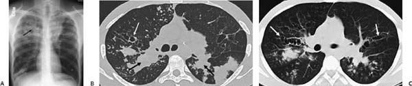
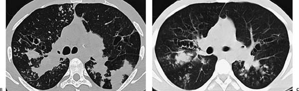
(A) Radiograph of the chest shows ill-defined, multifocal parenchymal opacities bilaterally that are more confluent in the upper lobes. Irregular, elongated tubular densities are better appreciated on the right side (arrow). (B, C) Computed tomography (CT) of the chest with lung window images reveals cylindric bronchiectasis (white arrows) and the irregular tubular structure radiating from the hilar region to the periphery of the right lung (black arrows). Areas of tree-in-bud (TIB) opacity in the periphery of both lungs are also identified
 Differential Diagnosis
Stay updated, free articles. Join our Telegram channel

Full access? Get Clinical Tree


