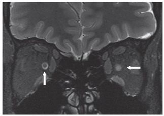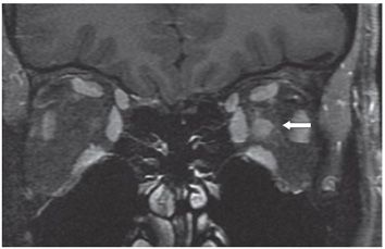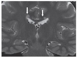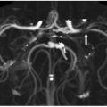


FINDINGS Figure 13-1. Coronal MR T1WI of the orbits. There is subtle asymmetry of the optic nerves (arrows). Figure 13-2. Coronal T2WI with fat suppression of the orbits through the same level as in Figure 13-1. There is hyperintensity of the left optic nerve with effacement of the surrounding cerebrospinal fluid (CSF) (transverse arrow). The right optic nerve is surrounded by hyperintense CSF within the sheath (vertical arrow). Figure 13-3. Coronal fat sat post-contrast T1WI through the orbits. There is contrast enhancement of the left optic nerve (arrow). Figure 13-4. Coronal T2WI through the corpus callosum. There are two corpus callosum hyperintense foci (arrows).
DIFFERENTIAL DIAGNOSIS Optic neuritis (ON), optic neuropathy, optic sheath meningioma, optic nerve glioma.
DIAGNOSIS Optic neuritis (ON) acute.
DISCUSSION
Stay updated, free articles. Join our Telegram channel

Full access? Get Clinical Tree


