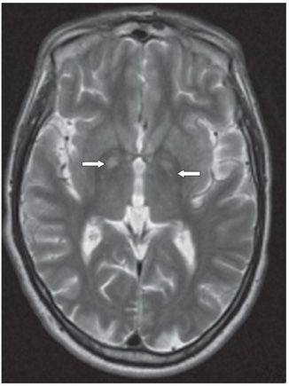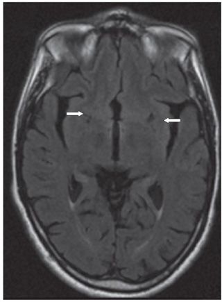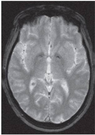


FINDINGS Figure 130-1. Axial NCCT is normal. Figure 130-2. Axial T2WI. There are bilateral hyperintense globus pallidus lesions (arrows). Figure 130-3. Axial FLAIR shows bilateral lentiform nuclei hypointensities (arrows). Figure 130-4. Axial GRE demonstrates bilateral symmetrical hyperintensity (arrows) with surrounding mineralization (hypointensities) in the medial lentiform nuclei.
DIFFERENTIAL DIAGNOSIS Hypertensive intracranial hemorrhage, anoxic infarcts, methanol intoxication, carbon monoxide poisoning, extrapontine osmotic demyelination syndrome, and Wilson disease.
DIAGNOSIS Methanol intoxication.
DISCUSSION
Stay updated, free articles. Join our Telegram channel

Full access? Get Clinical Tree


