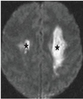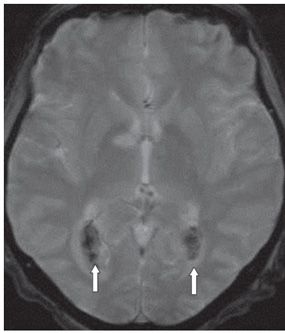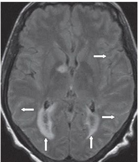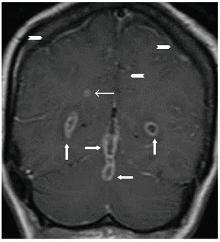


FINDINGS Figure 131-1. Axial DWI through the level of the frontal horns of the lateral ventricles. There is a left frontal white matter (WM) and a left peritrigonal focal restricted diffusion consistent with acute infarcts (vertical arrows). There is subtle multi-focal diffusion restriction in the splenium centrally (line arrow) and within the trigones and subarachnoid spaces. Figure 131-2. Axial DWI through the level of the centrum semiovale showing bilateral deep WM restricted diffusion (stars). Figure 131-3. Axial GRE through the trigones. There is blooming within the trigones (arrows) consistent with hemorrhage. Figure 131-4. Axial FLAIR through the trigones. There is hyperintensity within the convexity subarachnoid spaces (transverse arrows) suggesting meningitis or subarachnoid hemorrhage (SAH). There is bilateral peritrigonal confluent hyperintensity consistent with edema (vertical arrows) Figure 131-5
Stay updated, free articles. Join our Telegram channel

Full access? Get Clinical Tree


