CASE 132
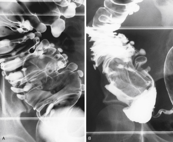
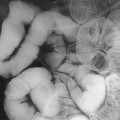
History: A 43-year-old woman underwent a CT scan for renal colic, and findings suspicious for a mass in the cecum were incidentally noted.
1. What should be included in the differential diagnosis of the imaging finding shown in Figure A? (Choose all that apply.)
A. Fatty infiltration of the ileocecal valve
B. Lipoma of the ileocecal valve
2. Which of the following statements regarding the ileocecal valve at barium enema is true?
A. The ileocecal valve is usually directly visible.
B. A lobulated mucosal surface is suspicious for malignancy.
C. Asymmetry of the valve lips indicates infiltration by disease.
D. Filling of the terminal ileum is required to identify the ileocecal valve.
3. Which of the following statements regarding disease at the ileocecal valve is true?
Stay updated, free articles. Join our Telegram channel

Full access? Get Clinical Tree


