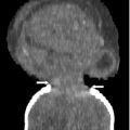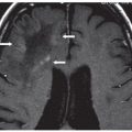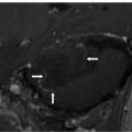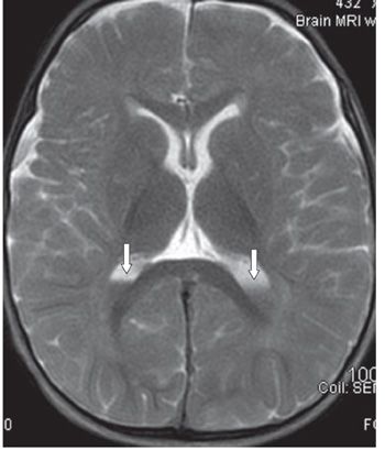
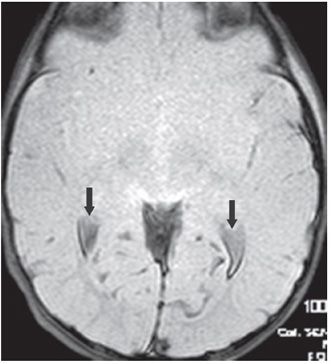
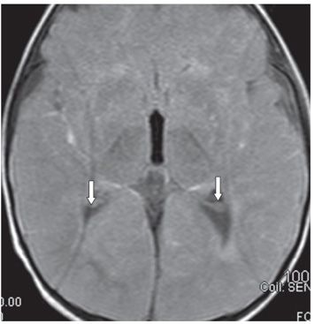
FINDINGS Figure 132-1. Axial NCCT through the lateral ventricles. There is bilateral intraventricular hyperdensity with blood–cerebrospinal fluid (CSF) levels in the trigones (arrows). There is diffuse brain hypodensity with no white–gray matter differentiation and effacement of convexity sulci consistent with brain edema and swelling. Figure 132-2. Axial T2WI through the lateral ventricles. There are fluid–fluid levels in the trigones (arrows) with hypointense dependent collections. Figure 132-3. Axial GRE through the lateral ventricles. There is bilateral intraventricular hypointensity with fluid–fluid levels in the trigones (arrows). Figure 132-4. Axial FLAIR through the lateral ventricles. There is bilateral intraventricular hypointensity with fluid–fluid levels in the trigones (arrows). The dependent hemorrhage in the trigones is slightly hyperintense compared with the CSF in the third ventricle. There is visible sulcal hyperintensities consistent with subarachnoid hemorrhage.
DIFFERENTIAL DIAGNOSIS Ventriculitis, intraventricular hemorrhage (IVH).
DIAGNOSIS Acute intraventricular hemorrhage (acute IVH) in acute hypoxic-ischemic encephalopathy.
DISCUSSION
Stay updated, free articles. Join our Telegram channel

Full access? Get Clinical Tree


