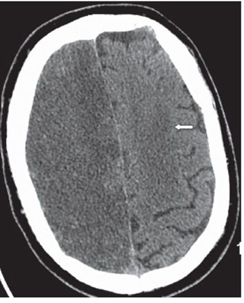
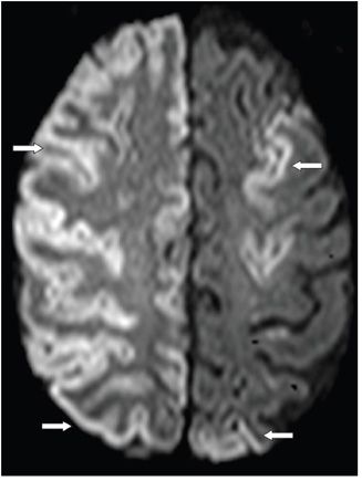
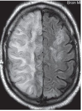
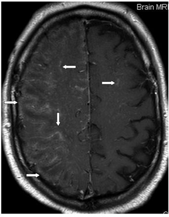
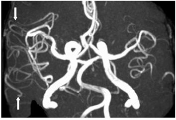
FINDINGS Figure 133-1. Axial NCCT through the centrum semiovale. There are multiple gas bubbles measuring up to −450 HU in the right cerebral parenchyma and within the right convexity sulci (arrows). Fewer similar gas bubbles are present in the left frontal lobe. Figure 133-2. Axial follow-up NCCT within 24 hours. There is resolution of the gas bubbles. There is diffuse right cerebral hemisphere hypodensity with no gray matter (GM)–white matter (WM) differentiation. There is effacement of the right convexity sulci. Similar but less prominent hypodensity is present in the left cerebral hemisphere (arrow). Figure 133-3. Axial DWI a few hours following Figure 133-2. There is extensive right hemispheric cortical ribbon hyperintensity consistent with cortical laminar necrosis (arrows). There is underlying WM smudgy hyperintensity. Similar but patchy changes are present in the left hemisphere. Figure 133-4
Stay updated, free articles. Join our Telegram channel

Full access? Get Clinical Tree


