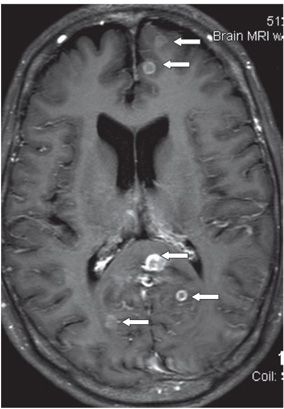
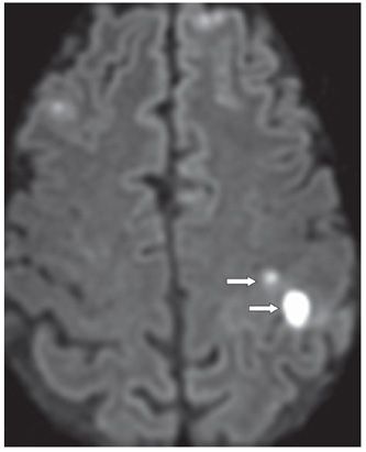
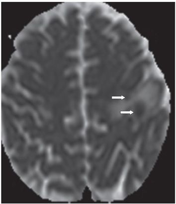
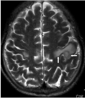
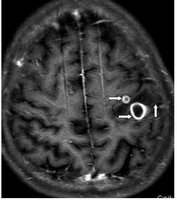
FINDINGS Figures 135-1 and 135-2. Axial FLAIR and post-contrast T1WI MRI through the splenium of the corpus callosum showing multiple ring-enhancing small lesions measuring about 1 cm or less in size in the left parasagittal frontal lobe, left splenium, and in bilateral occipital lobes (arrows). Lesions are surrounded by very minimal hyperintensity on the FLAIR except in areas where they coalesce as in the splenium where the surrounding T2 hyperintensity is large. Lesions are mostly cortical and/or subcortical in location. Figures 135-3, 135-4, 135-5, and 135-6. Axial DWI with corresponding ADC map, T2WI, and post-contrast T1WI through the superior centrum semiovale. There are more multifocal cortical subcortical lesions with central diffusion restriction in bilateral frontal lobes. The two posterior left frontal lobe lesions (transverse arrows) show smooth thick ring enhancement in Figure 135-6 with surrounding vasogenic edema (vertical arrows in figures 5 and 6). The larger left posterior frontal lobe lesion measures about 1.6 cm with a hyperintense core and hypointense rim on the T2WI (Figure 135-5
Stay updated, free articles. Join our Telegram channel

Full access? Get Clinical Tree


