CASE 136
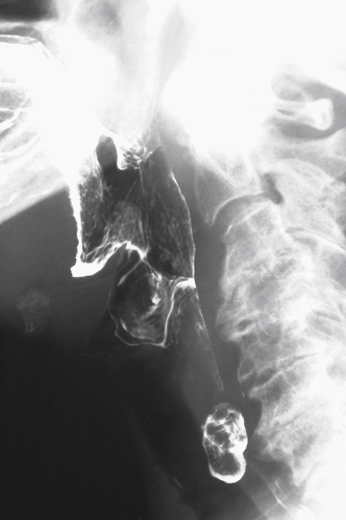
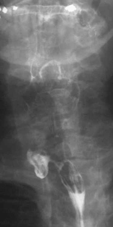
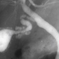
History: An 83-year-old woman presents with suprasternal dysphagia.
1. What should be included in the differential diagnosis of the imaging finding shown in the figures? (Choose all that apply.)
D. Killian-Jamieson diverticulum
E. Lateral hypopharyngeal pouch
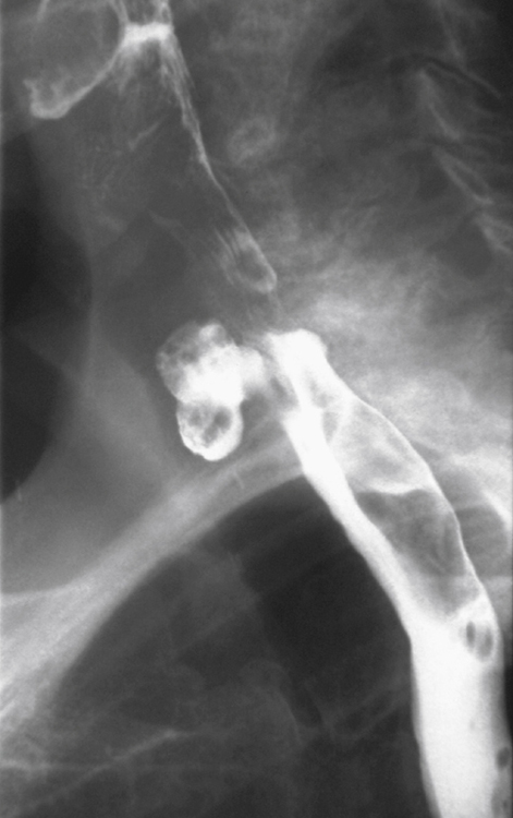
2. What is the anatomy of the site of weakness where a Killian-Jamieson diverticulum occurs?
B. Between the oblique and transverse fibers of the cricopharyngeus muscle
C. Between the inferior pharyngeal constrictor muscle and the thyrohyoid muscle
3. What specific complication is a patient at risk of with treatment of this lesion?
Stay updated, free articles. Join our Telegram channel

Full access? Get Clinical Tree


