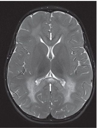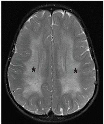

FINDINGS Figure 137-1. Axial DWI through the splenium of corpus callosum. There is hyperintensity of the splenium (arrow). The ADC map (not shown) is consistent with restricted diffusion. Figures 137-2 and 137-3. Axial T2WI through the splenium and centrum semiovale, respectively. There is symmetric confluent white matter (WM) (sometimes called “butterfly”) hyperintensity (stars) extending from the ventricular walls to the subcortical WM. The splenium and genu of the corpus callosum (arrows) are involved. Linear tubular and punctate hypointensities are present within the confluent WM changes consistent with the so-called tigroid pattern.
DIFFERENTIAL DIAGNOSIS Pelizaeus-Merzbacher, TORCH, periventricular leukomalacia, metachromatic leukodystrophy (MLD), Krabbe disease.
DIAGNOSIS Metachromatic leukodystrophy (MLD).
DISCUSSION
Stay updated, free articles. Join our Telegram channel

Full access? Get Clinical Tree


