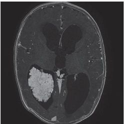
FINDINGS Figure 139-1. Axial post-contrast MR T1WI through the posterior fossa. There is an intensely enhancing rather knobbly or lumpy fourth ventricular mass surrounded by a thin rim of CSF. Figure 139-2. Axial post-contrast T1WI through the lateral ventricles in a different patient. There is a similar rather lumpy avidly contrast-enhancing mass with surrounding CSF within the right ventricular atrium.
DIFFERENTIAL DIAGNOSIS Choroid plexus papilloma, choroid plexus carcinoma, ependymoma, meningioma, medulloblastoma.
DIAGNOSIS Choroid plexus papilloma.
DISCUSSION
Stay updated, free articles. Join our Telegram channel

Full access? Get Clinical Tree


