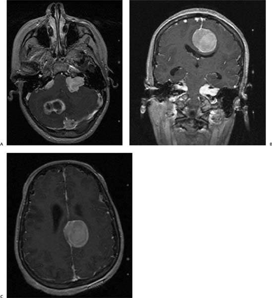
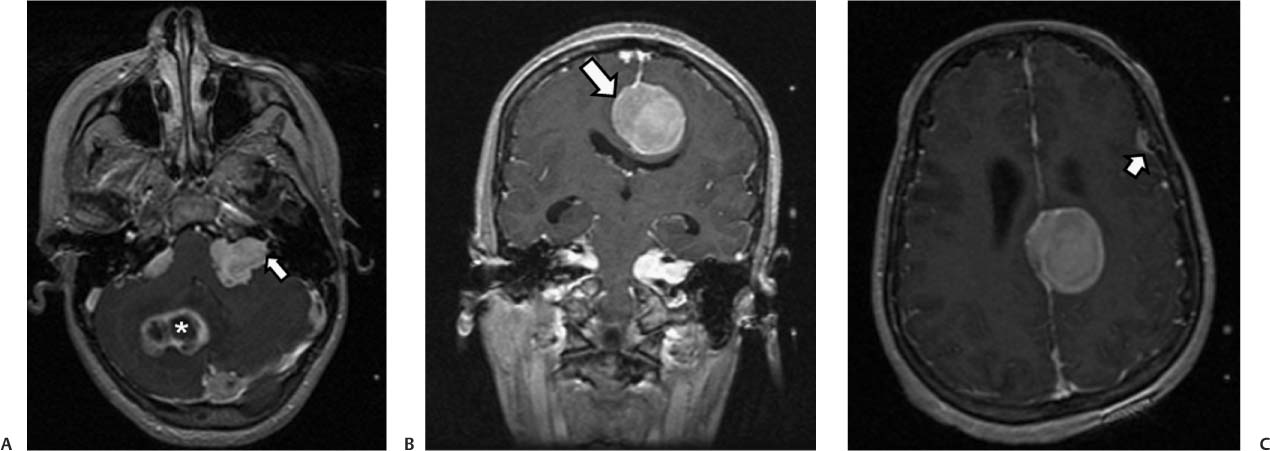
Case 14 A 22-year-old man presenting with hearing loss and tinnitus. (A) Axial postcontrast image of the posterior fossa demonstrates bilateral enhancing masses in the cerebellopontine angle (CPA) cistern. On the left side, an intracanalicular component is noted (arrow). Two additional enhancing masses with central areas of fluid signal (asterisk) near the torcular and in the right cerebellum showed dural attachment in the tentorium on other images. (B) Coronal postcontrast image confirms the presence of CPA masses with an intracanalicular component and shows an additional enhancing lesion in the left parafalcine region with broad attachment to the dura (arrow). (C) Axial postcontrast image reveals another tiny dura-based mass in the left frontal convexity (arrow).
Clinical Presentation
Imaging Findings
Stay updated, free articles. Join our Telegram channel

Full access? Get Clinical Tree


