 Clinical Presentation
A 34-year-old man with fever, productive cough, and malaise 1 week after being hospitalized for diabetic ketoacidosis.
Further Work-up
 Imaging Findings
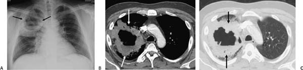
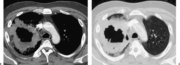
(A) Conventional radiograph of the chest shows a parenchymal opacity in the right upper lobe with a central area of cavitation (arrows). (B, C) Contrast-enhanced computed tomography of the chest confirms the presence of air-space consolidation in the posterior segment of the right upper lobe with central, thick-walled cavitation (arrows).
 Differential Diagnosis
• Klebsiella pneumonia: This Gram-negative bacillus, as well as other Gram-negative organisms (Escherichia coli and Pseudomonas aeruginosa), are important causes of nosocomial pneumonia and are more likely than other organisms to cause necrotizing disease and cavity formation.
• Tuberculosis:
Stay updated, free articles. Join our Telegram channel

Full access? Get Clinical Tree


