Case 14 The patient is a 58-year-old man with shortness of breath. (A) Image from a contrast-enhanced computed tomographic (CT) scan at the level of the pulmonary artery bifurcation demonstrating a large filling defect within the lumen of the right pulmonary artery (arrow). No other significant imaging findings are demonstrated. (B) Image of a selective right pulmonary arteriogram. There is a large filling defect within the superior segmental branch (white arrow
 Clinical Presentation
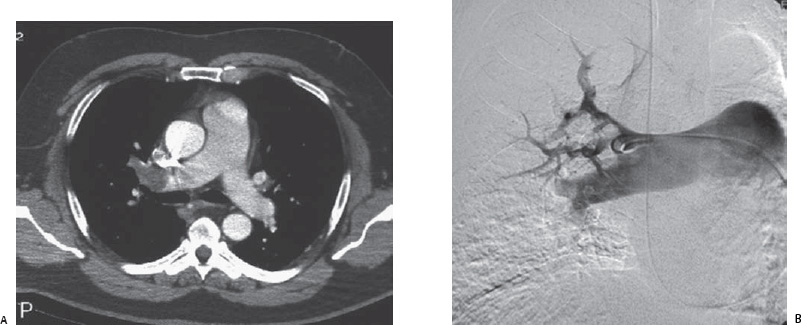
 Imaging Findings
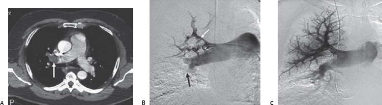
![]()
Stay updated, free articles. Join our Telegram channel

Full access? Get Clinical Tree


