Case 14
 Clinical Presentation
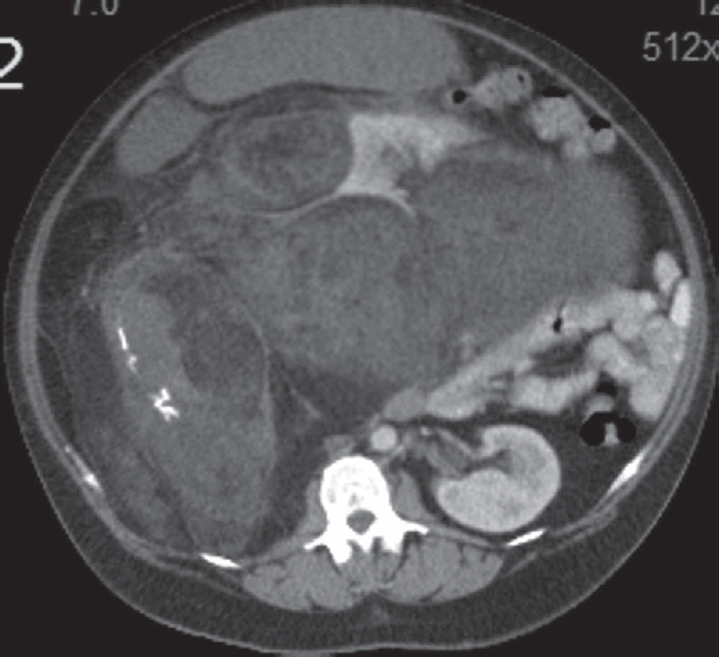
A 42-year-old woman who underwent computed tomography for increasing abdominal girth.
 Imaging Findings
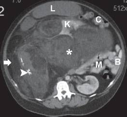
Contrast-enhanced computed tomography shows a large, bizarre-appearing mass in the posterior abdomen (arrow) that is displacing the right kidney (K), liver (L), ascending colon (C), small bowel (B), and mesentery (M). There is scalloping of the right renal outline, but no defect is seen in the renal parenchyma. The mass is composed predominantly of fat with large areas of soft tissue (asterisk). A few septa course through it. Some calcifications are present (arrowhead). No large vessels traverse the mass.
 Differential Diagnosis
• Retroperitoneal liposarcoma: All fat-containing tumors centered in the retroperitoneal adipose tissue should be considered liposarcoma unless proven otherwise. The soft-tissue elements contain areas of dedifferentiation and malignant change. Soft-tissue septa are also commonly seen. No large vessels course through retroperitoneal sarcomas.
• Retroperitoneal lipoma:
Stay updated, free articles. Join our Telegram channel

Full access? Get Clinical Tree


