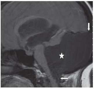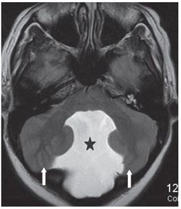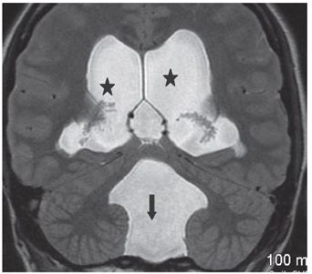


FINDINGS Figure 141-1. Axial NCCT through fourth ventricle. Large fourth ventricle ballooned into a large dorsal posterior fossa cyst (star) with remodeling of the occipital bone. The cerebellum is compressed anterolaterally (arrows). The third (chevron) and lateral ventricles (vertical arrows) are severely dilated. Figure 141-2. Sagittal MR T1WI through the fourth ventricle. There is a large posterior fossa cyst (star) compressing the brainstem and displacing the torcula upward (arrow). The cyst extends through the foramen magnum (transverse arrow). The third and lateral ventricles are dilated. Figure 141-3. Axial T2WI through the fourth ventricle. The fourth ventricle opens into a dorsal midline cyst (star). Cerebellum is rotated and compressed laterally (arrows). There is effacement of the subarachnoid space. Figure 141-4. Coronal T2WI through the fourth ventricle. The fourth ventricle opens into a dorsal midline cyst (arrow). Cerebellum is compressed laterally. The lateral ventricles are dilated (stars).
DIFFERENTIAL DIAGNOSIS Dandy-Walker malformation (DWM), mega cisterna magna (MCM), posterior fossa arachnoid cyst (PFAC).
DIAGNOSIS Dandy-Walker malformation (DWM).
DISCUSSION
Stay updated, free articles. Join our Telegram channel

Full access? Get Clinical Tree


