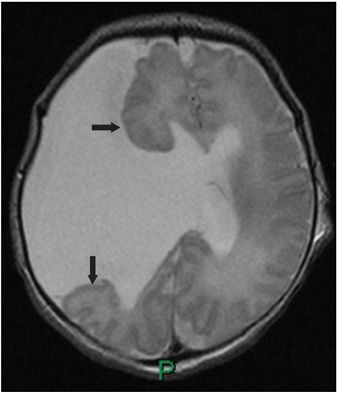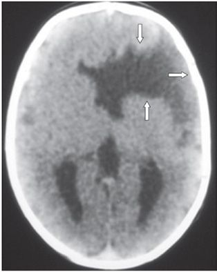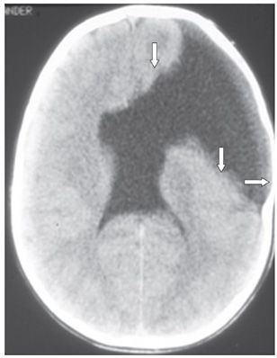


FINDINGS Figure 142-1. Axial NCCT through the lateral ventricles. There are bilateral suprasylvian coronal clefts lined by cortical gray matter (GM) (arrows). The clefts extend from the subarachnoid space to the lateral ventricular walls. This is consistent with “closed lip” schizencephaly. The ventricles are abnormally shaped with nonvisualization of the septum pellucidum. Figure 142-2. Axial T2WI through the lateral ventricles in a companion patient. There is a wide cleft through the right cerebral hemisphere linking the wide open lateral ventricle to the subarachnoid space. The margin of the cleft is lined by cortical GM (arrows). This is an open lip schizencephaly. The splenium of the corpus callosum and the septum pellucidum are absent. Figures 142-3 and 142-4. Axial NCCT through the lateral ventricles in another companion patient as a baby (Figure 142-3
Stay updated, free articles. Join our Telegram channel

Full access? Get Clinical Tree


