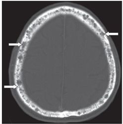
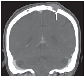
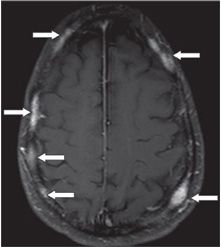
FINDINGS Figure 143-1. Skull film PA view. There are innumerable lucent lesions throughout the calvarium (arrows point to some of them). Figure 143-2. Axial NCCT through the vault. There are corresponding multiple hypodense cranial vault lesions, some well-defined, and all without sclerosis. Figure 143-3. Coronal reconstruction of CECT centered on one lesion with a prominent extraosseous subgaleal soft tissue component (arrow). Figure 143-4. Axial post-contrast T1WI. There are multiple contrast-enhancing scattered intradiploic lesions (arrows point to some of them).
DIFFERENTIAL DIAGNOSIS Paget disease, metastases, multiple myeloma, lymphoma.
DIAGNOSIS Multiple myeloma.
DISCUSSION


Stay updated, free articles. Join our Telegram channel

Full access? Get Clinical Tree


