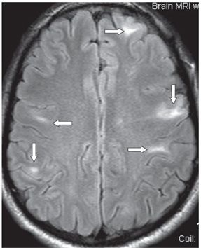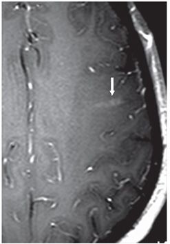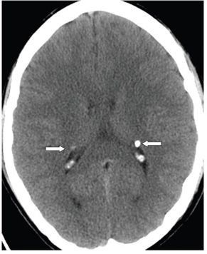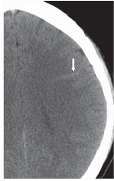


FINDINGS Figure 145-1. Axial GRE through the lateral ventricles. There is a left lateral ventricular subependymal round hypointensity (arrow). Figure 145-2. Axial FLAIR image through the level of the centrum semiovale showing multifocal radial white matter hyperintense bands radiating from the subcortical regions in bilateral cerebral hemispheres (arrows). Two left frontal lobe lesions show associated cortical thickening of intermediate intensity. Figure 145-3. Axial post-contrast T1WI through the level of the centrum semiovale. There is a left subcortical flame-shaped contrast enhancement (arrow). Figure 145-4. Axial NCCT through the lateral ventricles. There are bilateral subependymal hyperdensities consistent with calcifications (arrows). Figure 145-5. Axial NCCT through the left centrum semiovale. There is a thick linear left frontal subcortical hyperintensity consistent with a subcortical tuber (arrow).
DIFFERENTIAL DIAGNOSIS Toxoplasmosis, tuberous sclerosis complex (TSC).
Stay updated, free articles. Join our Telegram channel

Full access? Get Clinical Tree


