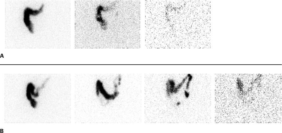
CASE 145 A 44-year-old woman presents with a history of chronic constipation that has become difficult to treat. The patient had an episode of diarrhea 24 hours following the initial set of images, and therefore the study was rescheduled and repeated several weeks later. Fig. 145.1 (A) Initial study. (B) Second study. • A 1 mCi dose of 111In-DTPA is ingested in 500 mL of water. • Anterior and posterior images of the abdomen are acquired at 6, 24, 48, and 72 hours after ingestion of the radiolabeled water. • Regions-of-interest are defined for the cecum, ascending colon, transverse colon, descending colon, and rectum, and for excreted stool. • The geometric mean of the anterior and posterior images is obtained for each imaging time point for each region. • All counts are corrected for decay. • The geometric center of activity (weighted average of the counts in each region) is determined at each imaging time for each region of the colon. The initial set of images (Fig. 145.1A) demonstrates accumulation of tracer in the ascending and trans-verse colon. After an episode of diarrhea, the diminished tracer can still be seen collecting just behind the mid–transverse colon. A second study (Fig. 145.1B), done several days later, again shows prominent accumulation of radiotracer in the transverse colon on 24-hour images. At 48 hours, some tracer is seen to move beyond the transverse colon to the left side of the colon. At 72 hours, much of the activity has been evacuated.
Clinical Presentation
Technique
Image Interpretation
Stay updated, free articles. Join our Telegram channel

Full access? Get Clinical Tree


