CASE 146
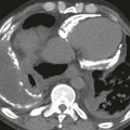
1. What etiologies can cause this finding? (Choose all that apply.)
D. Tuberculosis
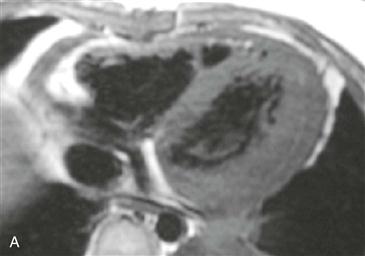
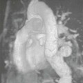
2. What is the pericardial abnormality in Fig. A?
A. Effusion
B. Thickening
D. Nodularity
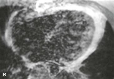
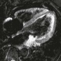
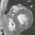
3. Given the symptoms and both images, what is the most likely diagnosis?
B. Inflammatory constrictive pericarditis
C. Effusive constrictive pericarditis
D. Tumor
Stay updated, free articles. Join our Telegram channel

Full access? Get Clinical Tree


