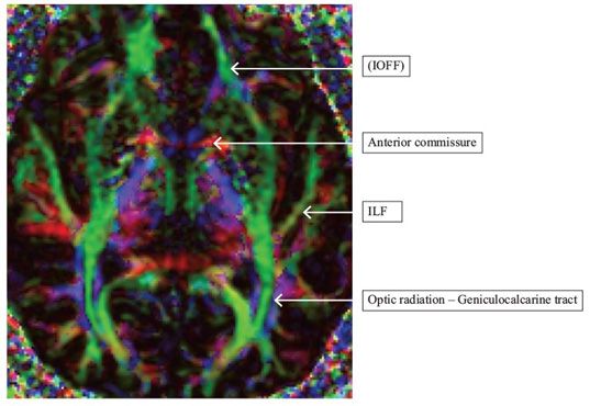
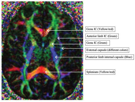
FIGURE 146-3 Basal ganglia level
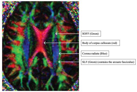
FIGURE 146-4 Body of corpus callosum level
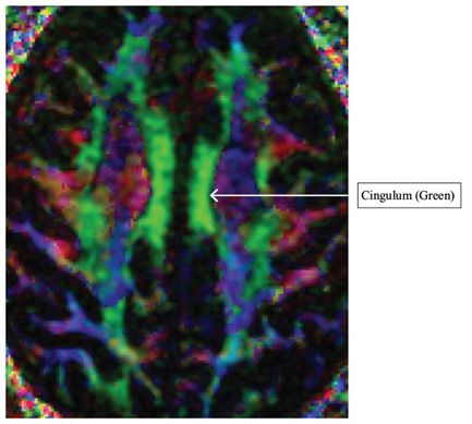
FIGURE 146-5 Cingulum level above corpus callosum
FINDINGS All images are axial DTI color directional maps. The color hue indicates direction of fiber tracts: green indicates anteroposterior direction; blue indicates craniocaudal or superoinferior direction; red indicates left to right direction.
Stay updated, free articles. Join our Telegram channel

Full access? Get Clinical Tree


