Case 149
Case History
A 55-year-old presents with right areolar ulcer and bloody nipple discharge. Patient was initially sent for breast sonogram because of areolar induration. He has had removal of two previous squamous cell carcinomas on his face.
Physical Examination
• right breast: the nipple areolar complex is replaced by a large ulceration; area also indurated and erythematous
• left breast: normal exam
• neck: large left supraclavicular mass
Mammogram
Mass
• margin: circumscribed
• shape: oval
• density: high density
Calcifications
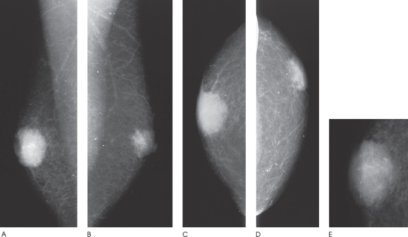
Figure 149–1. The sonographic mass corresponds to a right oval subareolar mammographic mass with heterogeneous calcifications. Although the mass appears circumscribed on the routine views (A, C), spot compression image (E) demonstrates that the mass has ill-defined margins. (A). Right MLO mammogram. (B). Left MLO mammogram. (C). Right CC mammogram. (D). Left CC mammogram. (E). Right CC spot compression mammogram.
Ultrasound


Stay updated, free articles. Join our Telegram channel

Full access? Get Clinical Tree


