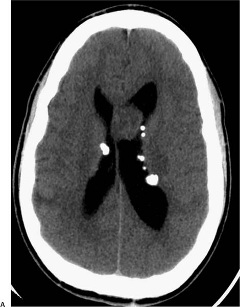
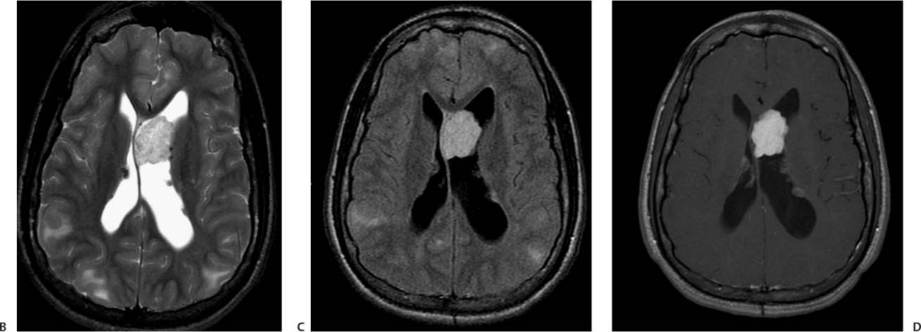
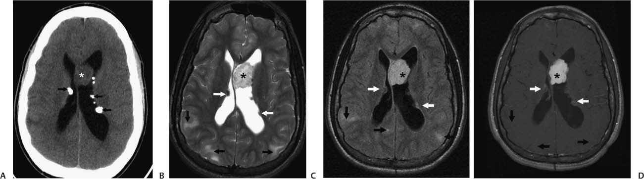
Case 15 A 19-year-old man with a history of seizures presenting with progressive mental status changes. (A) Axial computed tomography (CT) scan of the head without contrast shows multiple subependymal calcified nodules (arrows). A mass is seen in the left lateral ventricle adjacent to the septum pellucidum (asterisk). Note the dilatation of the left ventricle due to obstruction of the foramen of Monro on that side. (B) Axial T2-weighted image (WI) of the brain shows multiple subependymal nodules (white arrows). A giant cell astrocytoma with heterogeneous signal (asterisk) is deforming the septum pellucidum. Multiple subcortical and cortical tubers are seen as areas of hyperintensity (black arrows). (C) Fluid-attenuated inversion recovery (FLAIR) image of the brain shows multiple subependymal nodules (white arrows). A giant cell astrocytoma with heterogeneous signal (asterisk) is deforming the septum pellucidum. Multiple subcortical and cortical tubers are seen as areas of hyperintensity (black arrows). (D)
Clinical Presentation
Further Work-up
Imaging Findings
![]()
Stay updated, free articles. Join our Telegram channel

Full access? Get Clinical Tree


