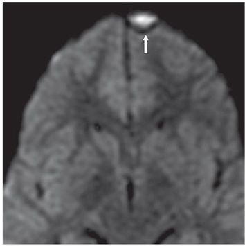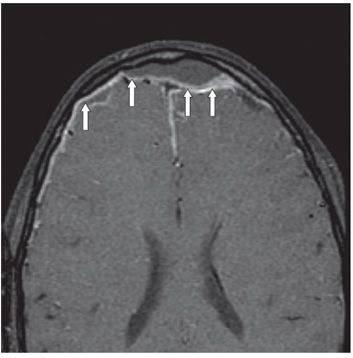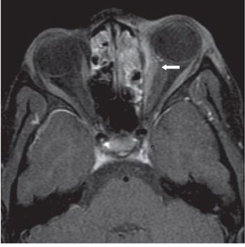


FINDINGS Figure 15-1. Axial T2WI through the inferior frontal lobes. There is hyperintense fluid opacification within both frontal sinuses (transverse arrows) with a small left frontal lenticular-shaped T2 hyperintense extraaxial collection (vertical arrow). Figure 15-2. Axial DWI demonstrates restricted diffusion within the left frontal extraaxial collection (arrow), indicating high cellularity within the collection and suggestive of an empyema. Figure 15-3. Axial post-contrast T1WI with fat saturation through the frontal lobes, performed 2 days after Figures 15-1 and 15-2. There is a much larger frontal epidural collection with thick dural enhancement and displacement (arrows). Note how the frontal epidural collection displaces the anterior falx posteriorly, confirming epidural rather than subdural location of the collection. Figure 15-4. Axial post-contrast T1WI with fat saturation through the ethmoid sinuses, performed at the same time. There is mucosal thickening and enhancement in the ethmoid air cells. There is a left orbital medial extraconal subperiosteal enhancement suggesting phlegmon (arrow). There is mild left orbital proptosis.
DIFFERENTIAL DIAGNOSIS Sinusitis with complication, leukemic or lymphomatous infiltration, epidural abscess.
DIAGNOSIS
Stay updated, free articles. Join our Telegram channel

Full access? Get Clinical Tree


