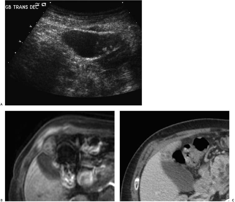
Case 15 A 53-year-old woman presents with vague abdominal pain. (A) Ultrasound shows a focal mass (arrowheads) in the fundus of the gallbladder (GB) with internal echogenic foci (arrows) without reverberation artifact. There is associated mural thickening. (B) Postinfusion T1 magnetic resonance imaging (MRI) shows the mass (arrow) to be focal and heterogeneous in signal intensity. The majority of the mass is similar to liver parenchyma except for central regions of low signal intensity. (C) Contrast-enhanced computed tomography (CT) shows the mass (arrow) to enhance similarly to liver parenchyma and to contain central, nonenhancing, cystic-appearing foci with a density similar to that of the GB lumen. • Focal adenomyomatosis: Given the GB wall thickening and an enhancing, masslike fundal structure with internal, nonenhancing (on CT and MRI), echogenic components, focal adenomyomatosis is the most likely diagnosis.

 Clinical Presentation
 Imaging Findings

 Differential Diagnosis
Stay updated, free articles. Join our Telegram channel

Full access? Get Clinical Tree


