CASE 15
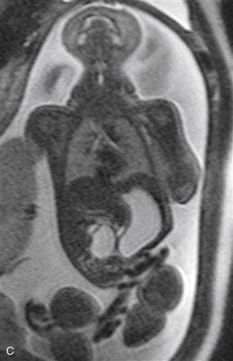
Used with permission from Anderson Publishing Ltd., from Victoria T, et al: Fetal MRI of common non-CNS abnormalities: a review. Appl Radiol 40[6]8-17, 2011. © Anderson Publishing Ltd.
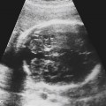
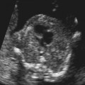
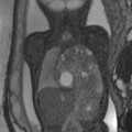
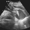
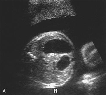
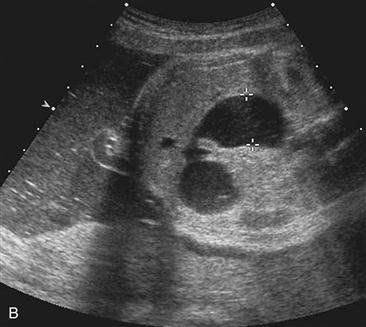
History: Axial scans through the upper abdomen in two separate fetuses with the same abnormalities are shown.
1. What should be included in the differential diagnosis of Figure A and Figure B? (Choose all that apply.)
C. Ovarian cyst
E. Renal cyst
2. The differential diagnosis of the “double bubble” sign of a dilated stomach and dilated duodenum would include all of the following except:
A. Malrotation with midgut volvulus
D. Hepatic cyst
3. Which of the following statements concerning duodenal atresia is false?
A. Approximately 40% of fetuses with trisomy 21 have duodenal atresia.
C. There is an increased risk of other intestinal atresia with duodenal atresia.
D. There is increased incidence of associated skeletal deformities with duodenal atresia.
Stay updated, free articles. Join our Telegram channel

Full access? Get Clinical Tree


