The Lenticulostriate Arteries and the Recurrent Artery of Heubner
15.1 Case Descriptions
15.1.1 Clinical Presentation
A 44-year-old male patient presented with acute subarachnoid hemorrhage.
15.1.2 Radiologic Studies
See ▶ Fig. 15.1, ▶ Fig. 15.2.
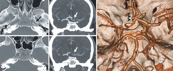
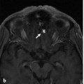
Fig. 15.1 CT in the bone window (a,b), CTA in coronal view (c,d), and 3D reconstruction (e) demonstrate hypoplasia of the left foramen lacerum and the carotid canal (black arrow), indicating congenital ICA hypoplasia. CTA confirms absence of intradural filling of the left ICA and provides evidence for reconstitution of the left MCA via two separate arteries arising from the anterior communicating complex (white arrows). As the source of hemorrhage, a small, broad-based anterior communicating artery aneurysm (black arrowheads) was found. Case is continued in ▶ Fig. 15.2.
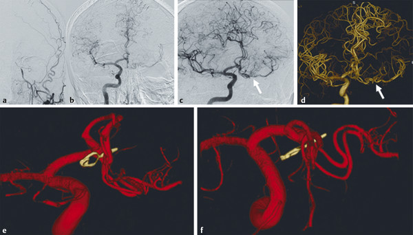
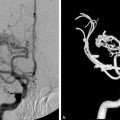
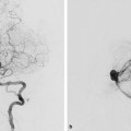
Fig. 15.2 After clip exclusion of the aneurysm, left common carotid artery angiogram in anteroposterior (AP) view (a) confirms ICA hypoplasia. Right ICA angiogram in AP (b), oblique (c), and 3D reconstruction (d,e,f) demonstrates reconstitution of the MCA via two arteries of the anterior communicating complex that run in parallel and from which the perforating lenticulostriate vessels (arrows) arise.
15.1.3 Diagnosis
Anterior communicating artery aneurysm in the setting of congenital hypoplasia of the left internal carotid artery (ICA) and reconstitution of the left middle cerebral artery (MCA) through two enlarged lenticulostriate arteries.
15.2 Embryology and Anatomy
The lenticulostriate arteries are a group of small perforating vessels arising from the proximal segments of the anterior cerebral artery (ACA) and the MCA to supply primarily, but not only, the striatum.
Embryologically, they develop from the lateral striate arteries, a system that arises from the rostral division of the internal cerebral artery, to meet the growing needs of the expanding telencephalic vesicles. This group of arteries will give rise to the lenticulostriate arteries, including the recurrent artery of Heubner (RAH), but will also form the adult MCA. Thus, from an embryological point of view, the MCA represents an enlarged perforator branch of the ACA. See also Case 14.
As discussed in the previous case, there is a broad range of variations related to the perforators as a result of differing coalescences during embryonic life for the different perforator groups; therefore, they may arise as multiple single small vessels from the parent artery or from a common larger trunk or take over each other’s territory. From medial to lateral, the following groups can be identified: the RAH, the medial lenticulostriate arteries from the ACA, the medial lenticulostriate arteries from the MCA, and the lateral lenticulostriate artery group. These groups can be interconnected with each other and form a rete (the most prominent example of which is the fenestration of the M1 or A1, as discussed in Case 14).
The RAH originates from the A1/A2 junction or in the first few millimeters of the A2 segment in 90% of cases. In only 10% of cases will it arise from the distal A1. The RAH supplies the anterior inferior striatum, anterior limb of the internal capsule, olfactory region, and anterior hypothalamus and is in a hemodynamic balance with the medial lenticulostriate arterial groups. Occasionally, the RAH can be duplicated or missing. The artery has a recurrent path turning laterally, coursing over the A1 (~60%), anterior to the A1 (~35%), and rarely, posterior to the A1 (3%). The territory supplied by the RAH is variable, as it is in balance with the medial lenticulostriate arteries (▶ Fig. 15.3; ▶ Fig. 15.4).
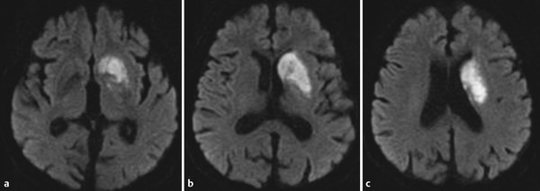
Fig. 15.3 Diffusion-weighted MRI (a,b,c) in a patient with an extensive left Heubner territory infarction demonstrates abnormal signal intensity within the head of the caudate nucleus and the anterior basal ganglia.
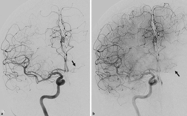
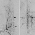
Fig. 15.4 Right ICA angiogram in AP view in arterial (a) and capillary (b) phases demonstrates the course and supply (capillary blush in b) of the left RAH (arrows).
Related posts:


Stay updated, free articles. Join our Telegram channel

Full access? Get Clinical Tree


