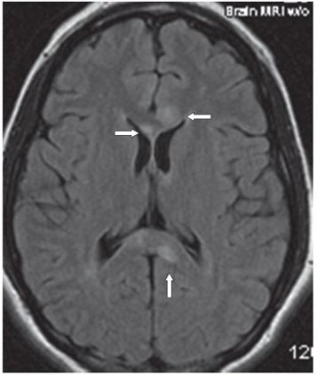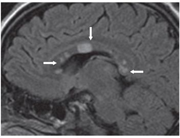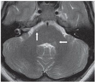


FINDINGS Figures 150-1 and 150-2. Axial DWI and FLAIR MRI through the corpus callosum (CC). There are multifocal CC, bilateral periventricular and subcortical hyperintense lesions with subtle lesions in the anterior thalamus bilaterally, and right cingulate cortex (arrows). Figure 150-3. Sagittal FLAIR MRI through the CC showing multiple round hyperintense lesions in the genu, body, and splenium of the CC (arrows) with no significant callososeptal interface hyperintensity. The so-called snowball lesion (vertical arrow) is a large round focal hyperintensity in the CC. Figure 150-4. Axial T2WI through the brachium pontis. There is a medial left brachium pontis focal hyperintensity adjacent to the fourth ventricle (transverse arrow) and a small lesion anteriorly in the basis pontis on the right (vertical arrow). There were no obvious areas of restricted diffusion on these images. Post-contrast images (not shown) did not reveal any abnormal contrast enhancement.
DIFFERENTIAL DIAGNOSIS Susac syndrome (SS), vasculitis, multiple sclerosis (MS), acute disseminated encephalomyelitis (ADEM).
DIAGNOSIS Susac syndrome (SS).
DISCUSSION
Stay updated, free articles. Join our Telegram channel

Full access? Get Clinical Tree


