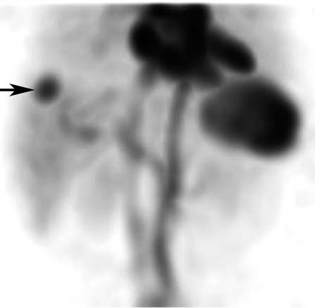
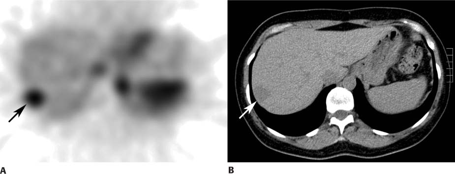
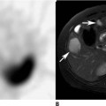
CASE 150 A 51-year-old woman with breast cancer undergoes abdominal ultrasonography to assess (arrow) for possible liver metastases. This reveals a 2.5-cm echogenic focus in the right lobe of the liver (Fig. 150.1). Although the sonographic appearance is suggestive of a hemangioma, it is not specific. Given the history of cancer, a tagged red blood cell (RBC) blood pool study is requested for confirmation (Figs. 150.2 and 150.3A). Fig. 150.1 Fig. 150.2 Fig. 150.3 • Three milliliters of autologous red blood cells are labeled with 99mTc-pertechnetate (25 mCi) and injected intravenously. • The patient is placed in a supine position beneath a gamma camera with a large field of view. • Flow images are acquired at 1 to 3 seconds per frame for 60 seconds (not shown). • Anterior abdominal image data are acquired continuously (1 minute per frame for 90 minutes). • Delayed dynamic images (1 minute per frame for 30 minutes) are acquired as needed. Anterior maximum-intensity projection (MIP) image (Fig. 150.2
Clinical Presentation

Technique
Image Interpretation
![]()
Stay updated, free articles. Join our Telegram channel


Full access? Get Clinical Tree


