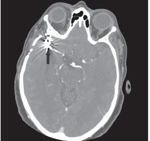
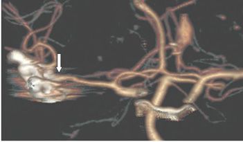
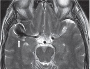
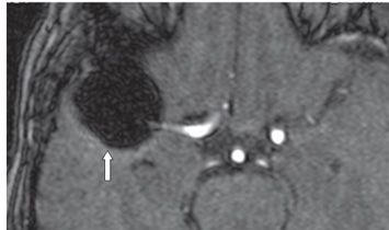
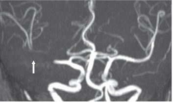
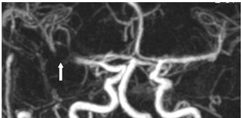
FINDINGS Figure 151-1. Lateral scout skull view for NCCT. The metallic clips in the paraclinoid region (transverse arrow) represent the aneurysm clips. The craniotomy bolts are present superiorly and anteriorly (vertical arrows). Figure 151-2. Axial CTA through the clips. There are significant streak artifacts through the clipped aneurysm rendering evaluation of completeness of clipping impossible. Figure 151-3. 3D CTA volume rendering of the aneurysm and clip. This offers the best way to show continuity of the vessel and the clipped aneurysm (arrow). But there is still question about how well the aneurysm has been obliterated. Figures 151-4 and 151-5


Stay updated, free articles. Join our Telegram channel

Full access? Get Clinical Tree


