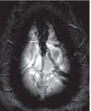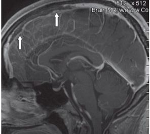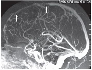


FINDINGS Figure 152-1. Axial FLAIR through the vertex. There are thick curvilinear hyperintense foci (arrows) in the posterior left frontal parasagittal convexity with associated effacement of left frontal sulci. Figure 152-2. Axial GRE through the superior sagittal sinus (SSS). There is enlarged blooming within the anterior SSS (arrow) and within the left vein of Trolard. Figure 152-3. Sagittal post-contrast T1WI. There is iso- to hypointense signal with peripheral enhancement in the anterior SSS (arrows). There is homogeneous opacification of the SSS posteriorly. Figure 152-4. 3D contrast-enhanced MRV. There is irregular severe narrowing of the anterior SSS (arrows) with smooth contrast opacification of the patent posterior SSS. There are multiple irregular collaterals over the frontal lobes.
DIFFERENTIAL DIAGNOSIS Subarachnoid hemorrhage (SAH), cortical infarcts, SSS thrombosis.
DIAGNOSIS SSS thrombosis (acute).
DISCUSSION
Stay updated, free articles. Join our Telegram channel

Full access? Get Clinical Tree


