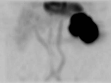
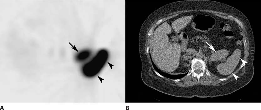
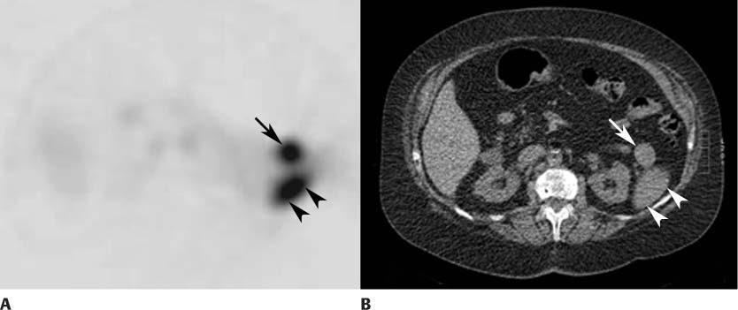
CASE 152 A 78-year-old woman undergoes chest radiography because of a cough. A potential lung nodule is identified, and a CT scan of the thorax is obtained. Images through the upper abdomen reveal two masses adjacent to the spleen. This leads to the study summarized below (Figs. 152.1, 152.2, and 152.3). Fig. 152.1 Fig. 152.2 Fig. 152.3 • Heat-damaged red blood cells (RBCs) labeled with 1 to 3 mCi of technetium 99mTc • Slow intravenous injection over 1 minute • Planar and SPECT images of the abdomen beginning 30 to 120 minutes after injection Anterior maximum-intensity projection (MIP) view from the SPECT study (Fig. 152.1) reveals intense uptake in the left upper quadrant. There is mild uptake in the cardiac blood pool and faint uptake in the liver. An axial view from the SPECT study (Fig. 152.2A
Clinical Presentation
Technique
 Autologous RBCs labeled in vitro
 Heated at 49°C for 15 to 20 minutes
 Cooled at room temperature for 15 minutes
Image Interpretation
![]()
Stay updated, free articles. Join our Telegram channel


Full access? Get Clinical Tree


