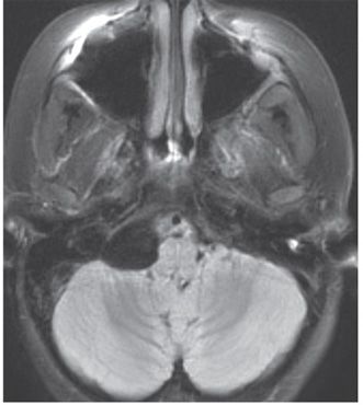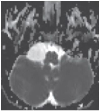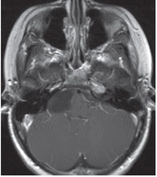


FINDINGS Figure 153-1. Axial T2WI through the posterior fossa at the level of cerebellopontine angles (CPAs). There is a well-circumscribed hyperintense extraaxial mass at the right CPA (star) causing mild mass effect upon the adjacent medulla. There is smooth erosion of the petrosal surface (arrow). Figure 153-2. Axial FLAIR through the mass shows complete signal suppression in the lesion, which is isointense to cerebrospinal fluid (CSF). Figure 153-3. ADC image shows high ADC without diffusion restriction. Figure 153-4. Axial post-contrast T1WI shows no appreciable enhancement in the lesion.
DIFFERENTIAL DIAGNOSIS Epidermoid cyst, neuroenteric cyst, arachnoid cyst, parasitic cyst, cystic neoplasm, that is, schwannoma, meningioma, or metastasis.
DIAGNOSIS CPA arachnoid cyst.
DISCUSSION
Stay updated, free articles. Join our Telegram channel

Full access? Get Clinical Tree


