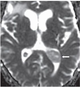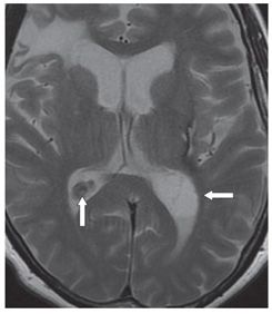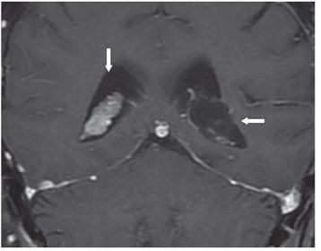


FINDINGS Figure 154-1. Axial DWI through the trigones. There is an ovoid hyperintense mass replacing the glomus of the left choroid plexus (arrow). Figure 154-2. ADC map through the trigones. There is intermediate hypointensity in the mass suggesting mildly restricted diffusion within the trigonal mass (arrow). Figure 154-3. T2WI through the trigone. The choroid plexus mass is hyperintense (arrow) hardly distinguishable from CSF. There are some linear hypointensity crisscrossing the mass. The right normal choroid plexus is hypointense (vertical arrow). Figure 154-4. Coronal post-contrast T1WI through the trigones. The left choroid plexus mass has similar intensity to CSF with a very thin rim of contrast enhancement (transverse arrow) presumably due to displaced choroid tissue. The right choroid plexus is avidly contrast enhancing as usual (vertical arrow).
DIFFERENTIAL DIAGNOSIS
Stay updated, free articles. Join our Telegram channel

Full access? Get Clinical Tree


