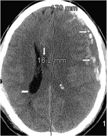
FINDINGS Figure 155-1. Axial NCCT through the basal ganglia. There is a left hemispheric mixed density (hyperacute) subdural hematoma (SDH) (transverse arrows). There is also some subarachnoid hemorrhage (SAH) beneath the SDH. The midline structures such as the septum pellucidum, third ventricle, and the calcified pineal gland (vertical arrows) have moved to the right of midline. The third and left lateral ventricles are compressed. Convexity sulci are effaced. There is mild dilation of the right lateral ventricle (chevron). Figure 155-2. Axial NCCT through the corona radiata. This demonstrates how to measure the shift; the septum pellucidum (vertical arrow) represents the midline structure measured against a line connecting the anterior and posterior attachment of the falx. The falx itself may bend away from the side of the mass or collection (transverse arrows point to the SDH). There is dilation of the right lateral ventricle (chevron).
DIFFERENTIAL DIAGNOSIS N/A.
DIAGNOSIS Subfalcine herniation (SFH) due to acute subdural hematoma.
DISCUSSION
Stay updated, free articles. Join our Telegram channel

Full access? Get Clinical Tree


