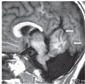
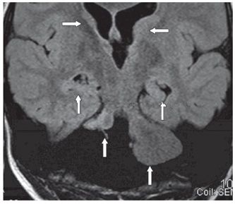
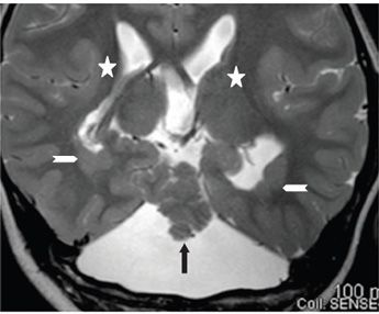
FINDINGS Figure 156-1. Sagittal T1WI through the posterior fossa. Small- to normal-sized posterior fossa. There is significantly reduced size of the cerebellum and vermis with a small remnant (chevron) and no recognizable cerebellar hemispheres. The fourth ventricle opens posteriorly inferiorly into a large cerebrospinal fluid (CSF) space. There is a fastigium (arrow). The tectum is thickened. The pons is small. The third ventricle (star) is large. The splenium is not formed. Figure 156-2. Left parasagittal T1WI. There is an oblique cleft through the occipital lobe (arrows). Figure 156-3


Stay updated, free articles. Join our Telegram channel

Full access? Get Clinical Tree


