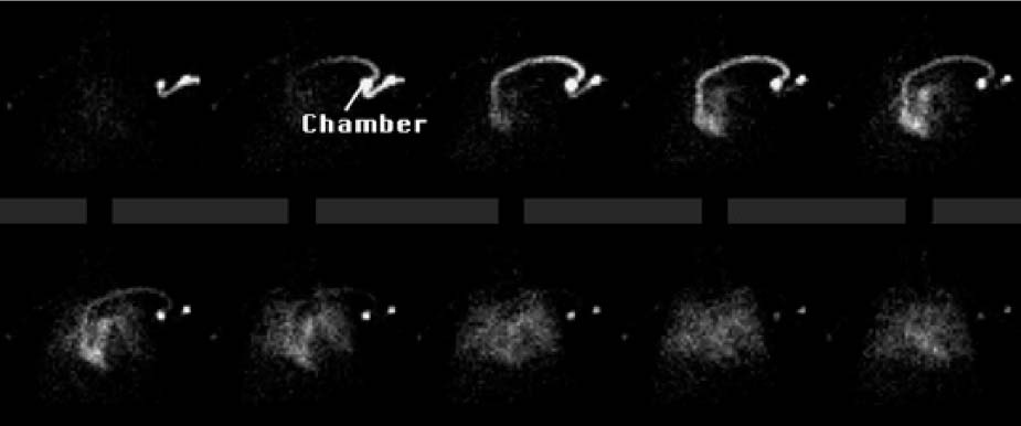
CASE 156 A 68-year-old woman presents with breast cancer and an indwelling left-sided central line (single-lumen Port-A-Cath; Smiths Medical, Dublin, OH). Blood return from the central line is poor. The study is performed to rule out obstruction or thrombus (Figs. 156.1, 156.2, and 156.3). Fig. 156.1 Fig. 156.2 Fig. 156.3 • To block thyroid uptake, potassium perchlorate (400 mg) is administered by mouth 15 minutes before99mTc-pertechnetate. • Indwelling catheters with subcutaneous chambers (such as those of the Port-A-Cath) need to be accessed with sterile technique. • With the patient lying supine, butterfly needles are attached to a three-way stopcock connected to the accessed central line ports and also placed into a vein in each upper extremity. • Connected to the stopcocks are 1-mL syringes containing approximately 5 mCi of 99mTc-pertechne-tate in a volume of 0.1 to 0.3 mL and 10-mL syringes containing 10 mL of normal saline. • Gamma camera with a large field of view • Low-energy, parallel-hole, high-resolution collimator • Energy window 20% centered at 140 keV • The camera is positioned over the anterior chest and shoulders with the heart in the center of the field of view. • The tracer is administered as a bolus injection, followed by the infusion of 10-mL flushes of normal saline in rapid sequence: first into the upper extremity ipsilateral to the central line, then the contralateral arm, and finally the central line. • Digital dynamic images are obtained in a 64 × 64 matrix at 1 frame per second for 120 seconds. At the completion of the study, the central line is flushed with 5 mL of heparin lock flush solution. • Data are reviewed in a cine format as well as 1 second per frame digital images.
Clinical Presentation
Technique
Image Interpretation
Stay updated, free articles. Join our Telegram channel

Full access? Get Clinical Tree


