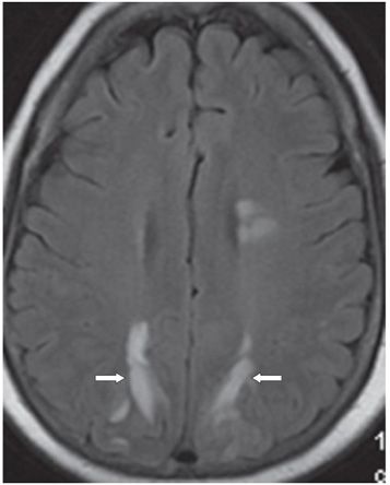
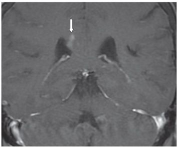
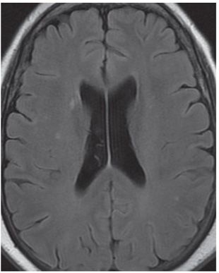
FINDINGS Figure 157-1. Axial T2 FLAIR through the lateral ventricles. There is bilateral parasagittal almost symmetrical parieto-occipital subcortical patchy hyperintensities. There is a collection of hyperintense signal in the splenium of the CC on the right (arrow). Figure 157-2. Axial T2 FLAIR through the high corona radiata. There is bilateral almost symmetrical parasagittal parietal subcortical hyperintensities (arrows). Additional left periventricular hyperintensities anteriorly. Figure 157-3. Coronal post-contrast T1WI through the splenium of the CC. There is focal contrast enhancement of the splenium on the right side (arrow) corresponding to the areas of FLAIR hyperintensity. Figure 157-4. Axial T2 FLAIR through the splenium 2 months after the initial study. There is resolution of the splenium and occipital/parietal lesions.
DIFFERENTIAL DIAGNOSIS Posterior reversible encephalopathy syndrome (PRES), acute disseminated encephalomyelitis (ADEM), metastases, lymphoma, cerebral venous thrombosis, gliomatosis cerebri, multiple watershed infarcts.
DIAGNOSIS PRES of CC.
DISCUSSION


Stay updated, free articles. Join our Telegram channel

Full access? Get Clinical Tree


