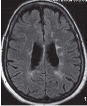
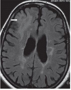
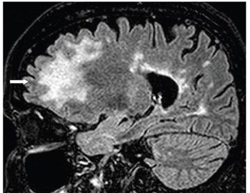
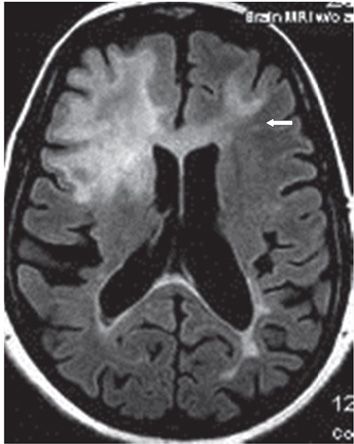
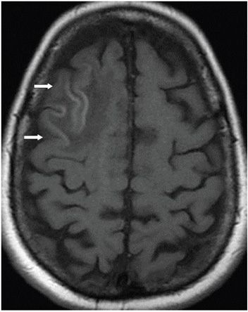
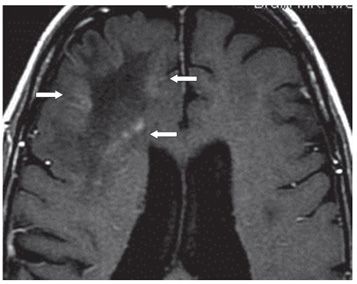
FINDINGS Figures 158-1 and 158-2. Right parasagittal and axial FLAIR MRI through the corona radiata, respectively. Baseline multifocal white matter (WM) hyperintensities of MS. Figures 158-3 and 158-4. Axial and right parasagittal FLAIR, respectively, through the corona radiata at the time of presentation with headache and memory loss. These show a large confluent right frontal mainly subcortical T2 hyperintense lesion without mass effect (arrows). Lesion was hypointense on T1WI and did not contrast enhance. Figure 158-5


Stay updated, free articles. Join our Telegram channel

Full access? Get Clinical Tree


