CASE 159
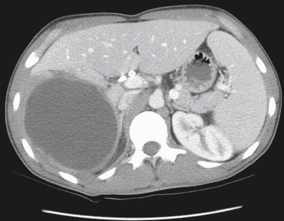
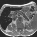
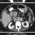
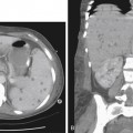
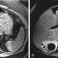
History: A 49-year-old man presents with right upper quadrant pain and a relevant past history of laparoscopic cholecystectomy 5 years ago.
1. What should be included in the differential diagnosis of the imaging finding shown in the figure? (Choose all that apply.)
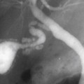
2. What imaging modality would you recommend to evaluate further for bile duct injury related to this lesion in planning for intervention?
A. Endoscopic retrograde cholangiopancreatography (ERCP)
C. MRI with magnetic resonance cholangiopancreatography (MRCP)
D. Percutaneous cholangiography
3. What is the most common complication of laparoscopic cholecystectomy?
Stay updated, free articles. Join our Telegram channel

Full access? Get Clinical Tree


