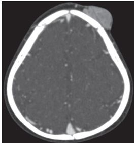
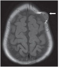
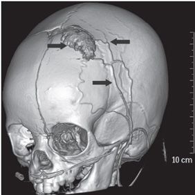
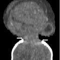
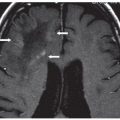
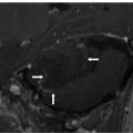
FINDINGS Figure 159-1. Coronal T2WI through posterior frontal bone. There is a left frontal scalp mass, mildly hyperintense to brain, with dark foci suggesting flow voids (arrow). Figure 159-2. Axial T1WI through the lesion. The mass is isointense to brain (arrow). Figure 159-3. Axial CTA source image. There is diffuse enhancement of the mass. The underlying bone appears intact. Figure 159-4. Volume rendered CTA. The lesion vascular supply is from branches of the external carotid artery (ECA) (arrows).
DIFFERENTIAL DIAGNOSIS Congenital hemangioma (CH), infantile hemangioma (IH), vascular malformation, hemangioendothelioma.
DIAGNOSIS IH.
DISCUSSION


Stay updated, free articles. Join our Telegram channel

Full access? Get Clinical Tree


