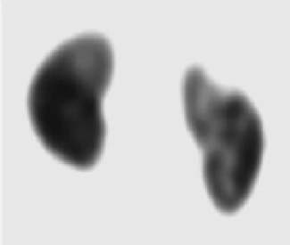
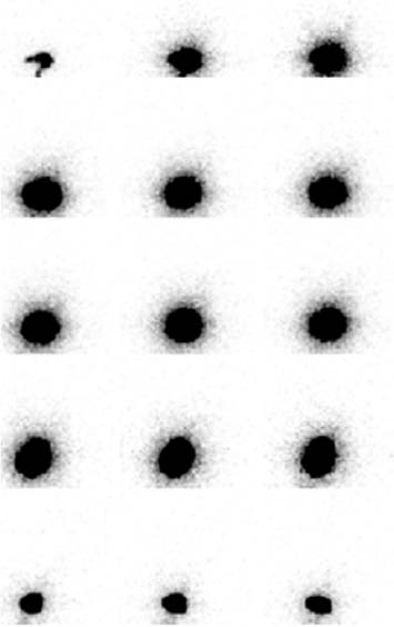
CASE 159 A 7-year-old girl presents with a 2-day history of abdominal pain, dysuria, and fever of 102° to 103°F. Physical examination shows right costovertebral angle tenderness. Initial laboratory evaluation includes a complete blood cell count (white blood cells, 13,200/mm3), urinalysis (white blood cells, 20–40 per high-power field; red blood cells, 10–20 per high-power field), blood culture, and urine culture. A 99mTc-DMSA study and a radionuclide cystography are requested for the evaluation of renal involvement and for the detection of vesicoureteral reflux. The patient was started on intravenous antibiotics for suspected pyelonephritis. Fig. 159.1 Fig. 159.2 • 99mTc-DMSA (0.5 mCi/kg) is given intravenously; the minimum dose is 0.2 mCi and the maximum dose is 3.0 mCi. • Use a high-resolution or ultra-high-resolution, low-energy, parallel-hole collimator for SPECT acquisition; perform planar imaging with a pinhole collimator for young infants. • Energy window is 20% centered at 140 keV. • Imaging is done 4 hours after radiotracer injection. SPECT acquisition is performed with 40 stops per detector or 120 stops with a three-detector imaging system. • 99mTc-pertechnetate (2 mCi) is given. The patient is asked to urinate before the examination. A catheter is placed into the bladder under sterile technique. A 500-mL bag of saline is connected to the catheter. The radiotracer is injected as a bolus into the catheter. The bladder is filled with saline at a pressure of 70 to 90 cm H2O. • A high-resolution, low-energy, parallel-hole collimator is used. • Energy window is 20% centered at 140 keV. • Dynamic images are acquired of the filling and voiding. Once the voiding is complete, the computer recording is terminated and the catheter is removed. A reprojected image of the kidneys (Fig. 159.1
Clinical Presentation
Technique
99mTc-DMSA Study
Radionuclide Cystography
Image Interpretation
![]()
Stay updated, free articles. Join our Telegram channel

Full access? Get Clinical Tree


