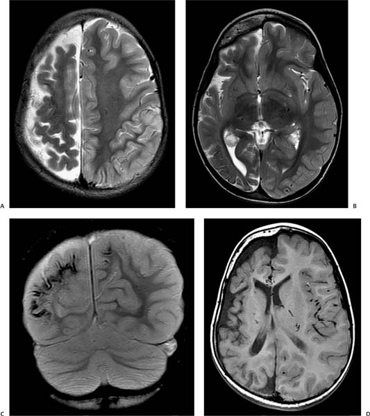
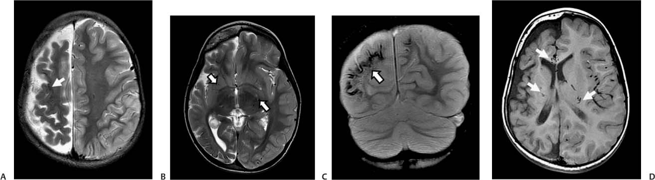
Case 16 A 17-year-old girl with seizures, hemiparesis, and homonymous hemianopsia. (A) Axial T2-weighted image (WI) of the brain demonstrates atrophy of the right cerebral hemisphere and linear areas of decreased signal along the cortex (arrow). (B) Axial T2WI of the brain demonstrates atrophy of the right cerebral hemisphere. Flow voids of dilated ependymal veins are also noted (arrows). (C) Coronal gradient-echo image reveals tramtrack cortical calcifications in the atrophic right hemisphere (arrow). Less extensive changes are present in the left parietal cortex. (D) Note the dilated medullary veins, which appear as large flow voids on the axial T1WI (arrows). • Sturge-Weber syndrome (also known as encephalotrigeminal angiomatosis):
Clinical Presentation
Imaging Findings
Differential Diagnosis
![]()
Stay updated, free articles. Join our Telegram channel

Full access? Get Clinical Tree


