 Clinical Presentation
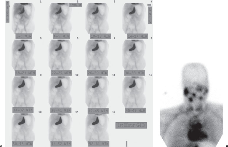
A patient with passage of bright red blood per rectum.
Further Work-up


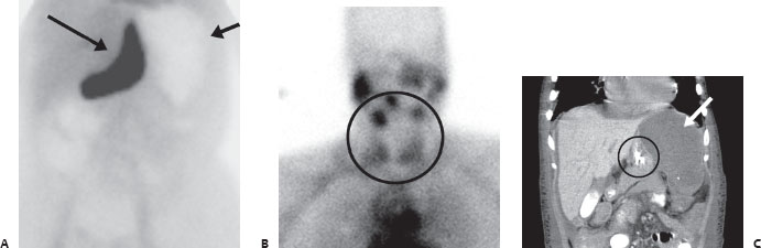
(A) Anterior images from Tc99m-tagged RBC study demonstrate immediate, intense, abnormal uptake in the upper mid abdomen (long arrow). After review of the CT scan, this localizes to the stomach, which is displaced medially by an enlarged, infarcted spleen. The infarcted spleen demonstrates no normal RBC accumulation (short arrow). (B) Anterior image of the neck reveals salivary and thyroid uptake, which is abnormal for an RBC scan (circle). (C) Coronal and axial enhanced abdominal CT scan shows the stomach (circle) displaced medially by an enlarged, hypodense, infarcted spleen (arrow).
 Differential Diagnosis
• Free pertechnetate artifact (and splenic infarct):


Stay updated, free articles. Join our Telegram channel

Full access? Get Clinical Tree


