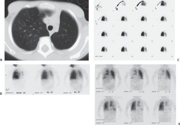
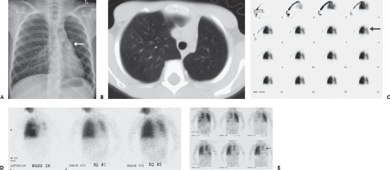
Case 16 A 10-year-old boy with cough. (A) Frontal chest radiograph demonstrates a small, hyperlucent left lung with diminished pulmonary vascularity. Left hilar vessels are present (arrow). (B) Axial computed tomography (CT) image confirms small left lung with decreased vascularity. (C) A nuclear medicine perfusion scan demonstrates markedly diminished perfusion of the left lung (arrow). (D,E) Nuclear medicine ventilation scans demonstrate decreased aeration of the left lung with air trapping (arrow). • Swyer-James syndrome:

Clinical Presentation
Further Work-up
Imaging Findings
Differential Diagnosis
![]()
Stay updated, free articles. Join our Telegram channel

Full access? Get Clinical Tree


