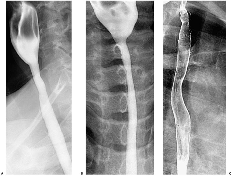
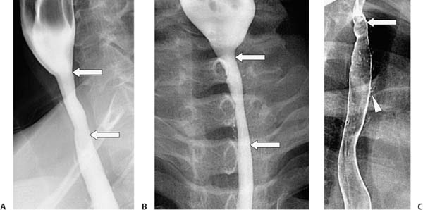
Case 16 A 52-year-old woman presents with heartburn and dysphagia. (A,B) Single-contrast esophagograms show circumfer ential narrowing (arrows) of the cervical esophagus with a smooth transition to normal-caliber esophagus. This finding persists on both left anterior oblique (A) and frontal (B) projections. (C) Double-contrast study again shows narrowing (arrow) of the cervical esophagus. Multiple small outpouchings (arrowhead) of barium in the proximal esophagus are consistent with intramural pseudodiverticulosis. • Barrett stricture: This classically occurs in the middle to upper esophagus and may be associated with intramural pseudodiverticulosis, as seen in this case. • Radiation injury from mediastinal irradiation: This occurs in the distribution of the radiation port. Patients with upper lobe, high mediastinal, or cervical malignancy may be at risk for this complication. • Skin diseases: Epidermolysis bullosa or benign pemphigoid may cause middle to upper strictures.
 Clinical Presentation
 Imaging Findings
 Differential Diagnosis
Stay updated, free articles. Join our Telegram channel

Full access? Get Clinical Tree


