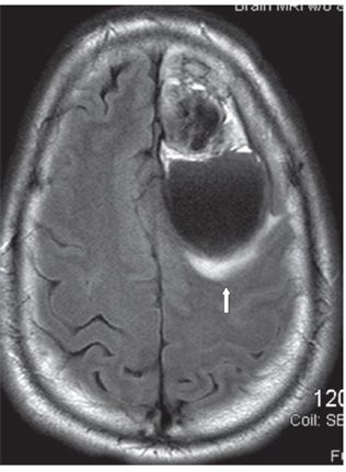
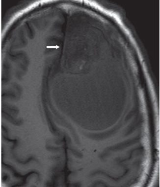
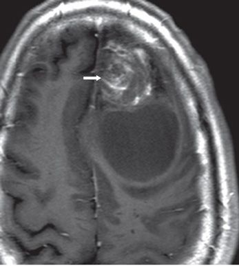
FINDINGS Figure 160-1. Axial NCCT through the frontal lobes. There is a large left frontal lobe intraaxial mass with an anterior hyperdense nodule (transverse arrow). There is a larger posterior cystic component with a hyperdense fluid component and level (vertical arrows). The hyperdense areas are consistent with hemorrhage. There is substantial mass effect with subfalcine herniation to the right (line arrow). Figure 160-2. Axial FLAIR through the centrum semiovale. The anterior nodule is predominantly hypointense with surrounding heterogeneous hyperintensity. The posterior cystic cavity is hypointense with surrounding hyperintensity—edema (arrow). Figures 160-3 and 160-4. Axial pre- and post-contrast T1WI. The anterior nodule is hypointense pre-contrast with heterogeneous contrast enhancement (arrows). The cystic posterior component does not enhance with contrast.
DIFFERENTIAL DIAGNOSIS


Stay updated, free articles. Join our Telegram channel

Full access? Get Clinical Tree


