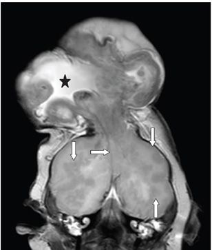
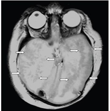
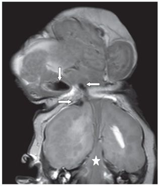
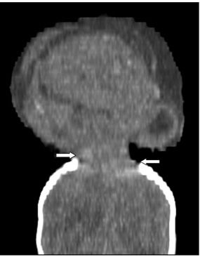
FINDINGS Figure 161-1. Sagittal T1WI through the vertex mass. Brain tissue along with meninges and cerebrospinal fluid (CSF) are seen protruding through a defect in the frontoparietal junction. The mass measures 8.5 cm × 7.1 cm × 5.3 cm. There is a linear hyperintense structure extending from the suprasellar cistern into the mass (anterior arrow) possibly a vascular pedicle. It appears as if the midline structures—ventricles, corpus callosum (posterior arrow)—have been flushed up through the defect. Figure 161-2. Coronal T2WI through the mass. There is dysgenetic brain, CSF, and meninges up in the mass. The large CSF space (star) probably represents the ventricles. The falx or a vascular pedicle (transverse arrow) extends into the mass. There are multiple heterotopic gray matter (GM) throughout the brain within the intracranial cavity (vertical arrows). Figure 161-3


Stay updated, free articles. Join our Telegram channel

Full access? Get Clinical Tree


