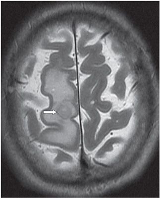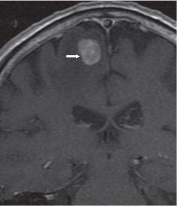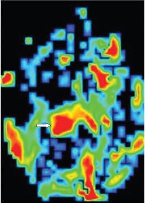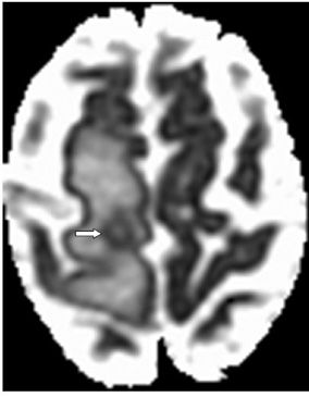


FINDINGS Figure 162-1. Axial non-contrast T1WI through the cerebral convexities. There is a round right posterior frontal lobe parasagittal small well-circumscribed mass (transverse arrow) in the premotor area, mildly heterogeneous but isointense compared to surrounding expanded hypointense white matter (WM) that is consistent with edema (vertical arrow). Figure 162-2. Corresponding axial T2WI. The mass is mildly heterogeneous and slightly hypointense (arrow) compared to the surrounding hyperintense edema. The right superior frontal gyrus is expanded, and adjacent sulci are partially effaced. Figure 162-3. Coronal post-contrast T1WI. There is a single mildly heterogeneous contrast-enhancing, sharply marginated, subcortical mass in the posterior parasagittal right frontal lobe. There is surrounding hypointensity consistent with edema or infiltrating tumor. Figure 162-4. Axial blood volume perfusion map through the mass. There is significant increase in the relative Cerebral Blood Volume (rCBV) (arrow). Figure 162-5
Stay updated, free articles. Join our Telegram channel

Full access? Get Clinical Tree


